T and B lymphocytes are central to immune defenses against infections and cancer but also drive autoimmune diseases (e.g., rheumatoid arthritis, SLE, multiple sclerosis), inflammatory disorders (colitis, psoriasis), and allergies/asthma. Ion channels and transporters (ICTs) sit at the heart of immune cell biology, regulating fundamental processes including growth, differentiation, metabolism, and gene expression. Beyond classical ions such as calcium, zinc, sodium, and chloride, ICTs also transport small molecules including metabolites, amino acids, and immune mediators. Despite the existence of more than one thousand ICTs, only 15–20 have been established as regulators of immune function — among them the calcium channel ORAI1 and the potassium channels Kv1.3 and KCa3.1, which are essential for T cell-mediated immunity.
Many fundamental questions remain unanswered: Which ICTs are expressed and functional across different lymphoid and myeloid cell types? What downstream signaling, metabolic, and epigenetic pathways do they engage? How do ICTs orchestrate complex immune responses to infection, tumors, or self-antigens? And can targeting immune cell ion channels lead to safe and effective treatments for autoimmune disease, allergy, or cancer?
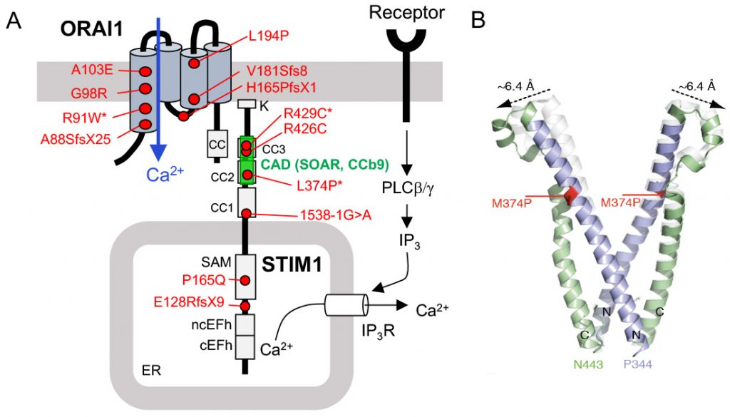
My laboratory addresses these questions through mechanistic and physiological studies in human and mouse immune cells. Our work builds on the landmark discovery of ORAI1 as the gene encoding the calcium release-activated calcium (CRAC) channel and the identification of CRAC channelopathy — a novel inborn error of immunity caused by mutations in ORAI1 and its activator STIM1. We have since uncovered several additional ICTs that regulate T and B cell function, including the chloride and cGAMP channel LRRC8C, multiple members of the ZIP family of zinc transporters, the sodium/bicarbonate cotransporter NBCn1/SLC4A7, and the copper transporter CTR1/SLC31A1.
Our research aims to discover and characterize novel immune ICTs, elucidate their molecular and immunological mechanisms, and ultimately lay the groundwork for an entirely new class of immunomodulatory therapies.
Overview
T and B lymphocytes are central to immune defenses against infections and cancer but also drive autoimmune diseases (e.g., rheumatoid arthritis, SLE, multiple sclerosis), inflammatory disorders (colitis, psoriasis), and allergies/asthma. Ion channels and transporters (ICTs) sit at the heart of immune cell biology, regulating fundamental processes including growth, differentiation, metabolism, and gene expression. Beyond classical ions such as calcium, zinc, sodium, and chloride, ICTs also transport small molecules including metabolites, amino acids, and immune mediators. Despite the existence of more than one thousand ICTs, only 15–20 have been established as regulators of immune function — among them the calcium channel ORAI1 and the potassium channels Kv1.3 and KCa3.1, which are essential for T cell-mediated immunity.
Many fundamental questions remain unanswered: Which ICTs are expressed and functional across different lymphoid and myeloid cell types? What downstream signaling, metabolic, and epigenetic pathways do they engage? How do ICTs orchestrate complex immune responses to infection, tumors, or self-antigens? And can targeting immune cell ion channels lead to safe and effective treatments for autoimmune disease, allergy, or cancer?
My laboratory addresses these questions through mechanistic and physiological studies in human and mouse immune cells. Our work builds on the landmark discovery of ORAI1 as the gene encoding the calcium release-activated calcium (CRAC) channel and the identification of CRAC channelopathy — a novel inborn error of immunity caused by mutations in ORAI1 and its activator STIM1. We have since uncovered several additional ICTs that regulate T and B cell function, including the chloride and cGAMP channel LRRC8C, multiple members of the ZIP family of zinc transporters, the sodium/bicarbonate cotransporter NBCn1/SLC4A7, and the copper transporter CTR1/SLC31A1.
Our research aims to discover and characterize novel immune ICTs, elucidate their molecular and immunological mechanisms, and ultimately lay the groundwork for an entirely new class of immunomodulatory therapies.
What are the Physiological Roles of Calcium Channels in Immunity?
Our laboratory has made seminal contributions to understanding the CRAC channel, beginning with the landmark discovery of ORAI1 as its pore-forming subunit. Leveraging genetically engineered mouse models with targeted deletion of ORAI1 and its homologues ORAI2 and ORAI3, as well as their activators STIM1 and STIM2, we have systematically dissected how CRAC channel-mediated calcium signaling governs immune cell physiology. Our studies revealed that CRAC channels control the expression of hundreds of genes in T cells, including cytokines and lineage-defining transcription factors that direct the differentiation of CD4⁺ T cells into distinct effector subsets, including Th1, Th2, Th17, and regulatory T (Treg) cells. In addition, CRAC channels drive key metabolic programs — encompassing glycolysis, mitochondrial respiration, and lipid metabolism — that are required to sustain T cell proliferation and proinflammatory effector functions. Collectively, these studies established ORAI and STIM proteins as the core molecular components of the CRAC channel and defined their essential roles in adaptive immunity.
Building on this foundation, we are investigating the molecular and immunological mechanism how CRAC channel-mediated calcium signals promote the differentiation of CD4⁺ T cells into T follicular helper (Tfh) cells, which are critical orchestrators of humoral immune responses in the context of infection and autoimmunity. In parallel, we are exploring how intracellular calcium homeostasis, governed by organellar calcium handling proteins, regulates B cell development into antibody-secreting plasma cells. Together, these studies provide fundamental insights into calcium signaling in lymphocyte biology and identify novel therapeutic targets for modulating humoral immunity in disease.
How do CRAC Channels control Immunity to Infection and Autoimmunity
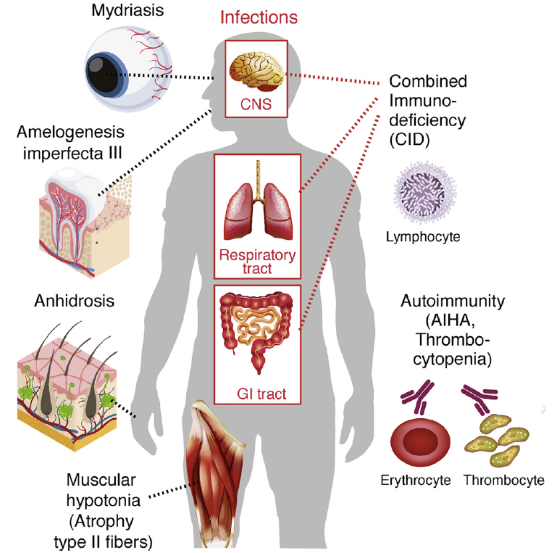
We identified the first patients with loss-of-function mutations in ORAI1 or STIM1, describing a new syndrome — CRAC channelopathy — characterized by combined immunodeficiency with recurrent infections, autoimmunity, muscular hypotonia, and ectodermal dysplasia. The severe susceptibility to infection in these patients underscores the non-redundant role of CRAC channel-mediated calcium signaling in adaptive immunity and provides a compelling human model for understanding how SOCE shapes immune defense. Using mice with genetic deletion of ORAI and STIM homologues, we demonstrated that CRAC channels are essential for cytotoxic CD8⁺ T cell responses to viral infection, CD4⁺ Th1 and T follicular helper (Tfh) cell responses to viral infection, and CD4⁺ T cell immunity to fungal and chronic mycobacterial infections — collectively providing a mechanistic basis for the immunodeficiency observed in CRAC channelopathy. Notably, the same pathway drives CD8⁺ T cell-mediated antitumor immunity across multiple cancer models.
CRAC channels also play essential roles in immune tolerance by controlling the differentiation and function of proinflammatory CD4⁺ T cell subsets. In animal models of inflammatory bowel disease, psoriasis, and multiple sclerosis, genetic deletion or pharmacological inhibition of CRAC channels attenuates Th1 and Th17 responses and reduces disease activity. CRAC channels similarly drive allergic asthma by regulating cell cycle progression and Th2 cell-defining transcription factors and cytokines. These pathogenic roles of are counterbalanced by CRAC channel function in regulatory T (Treg) cells, where calcium signals are required for thymic Treg development and the differentiation of tissue-resident Tregs and T follicular regulatory cells. Mice lacking CRAC channels in Tregs develop multiorgan autoimmunity resembling Sjögren’s disease, driven by elevated IFN-γ production. Together, our findings define a therapeutic window in which partial CRAC channel inhibition suppresses pathogenic inflammation while preserving immune tolerance and host defense.
Ongoing work in the lab continues to explore the roles of CRAC channels in infection, asthma, and autoimmune diseases including Sjögren’s disease and lupus.
Characterization of Novel Ion Channels and Transporters in Immune Function.
A major recent focus of my laboratory is the discovery and characterization of novel ion channels and transporters (ICTs) that regulate the function of T cells, B cells, and macrophages across diverse immune responses. Combining expression analyses of ICTs in human and mouse immune cell subsets with shRNA- and CRISPR/Cas9-based forward genetic screens, we have identified >10 new ICTs that govern multiple aspects of immune cell function in the context of infection, antitumor immunity, and autoimmunity. In CD4⁺ T cells, we identified the volume-regulated chloride channel LRRC8C and demonstrated that it transports the dinucleotide cGAMP to activate STING signaling, thereby controlling T cell expansion and immune responses. We further showed that CLNS1A — originally reported as a chloride channel subunit — is essential for the proinflammatory function of Th17 cells in models of multiple sclerosis and inflammatory bowel disease, acting as a cofactor of the protein arginine methyltransferase PRMT5 to regulate DNA repair. Additionally, the copper transporter SLC31A1 was found to regulate CD4⁺ T cell metabolism and pathogenic Th17 cell function, with consequent effects on autoimmunity. In B cells, we identified the amino acid transporter SLC7A5 and the bicarbonate transporter SLC4A7 as regulators of B cell metabolism through mTORC1, controlling plasma cell differentiation and antibody production.
Ongoing work in the laboratory is focused on elucidating the mechanisms by which ZIP family zinc transporters, SLC31A1, Ca2+ and Na+ transporters, and other ICTs govern T and B cell function and immune responses. The ultimate goal of these studies is to identify and validate novel therapeutic targets for modulating immunity in autoimmunity, cancer, and inflammatory disease.